
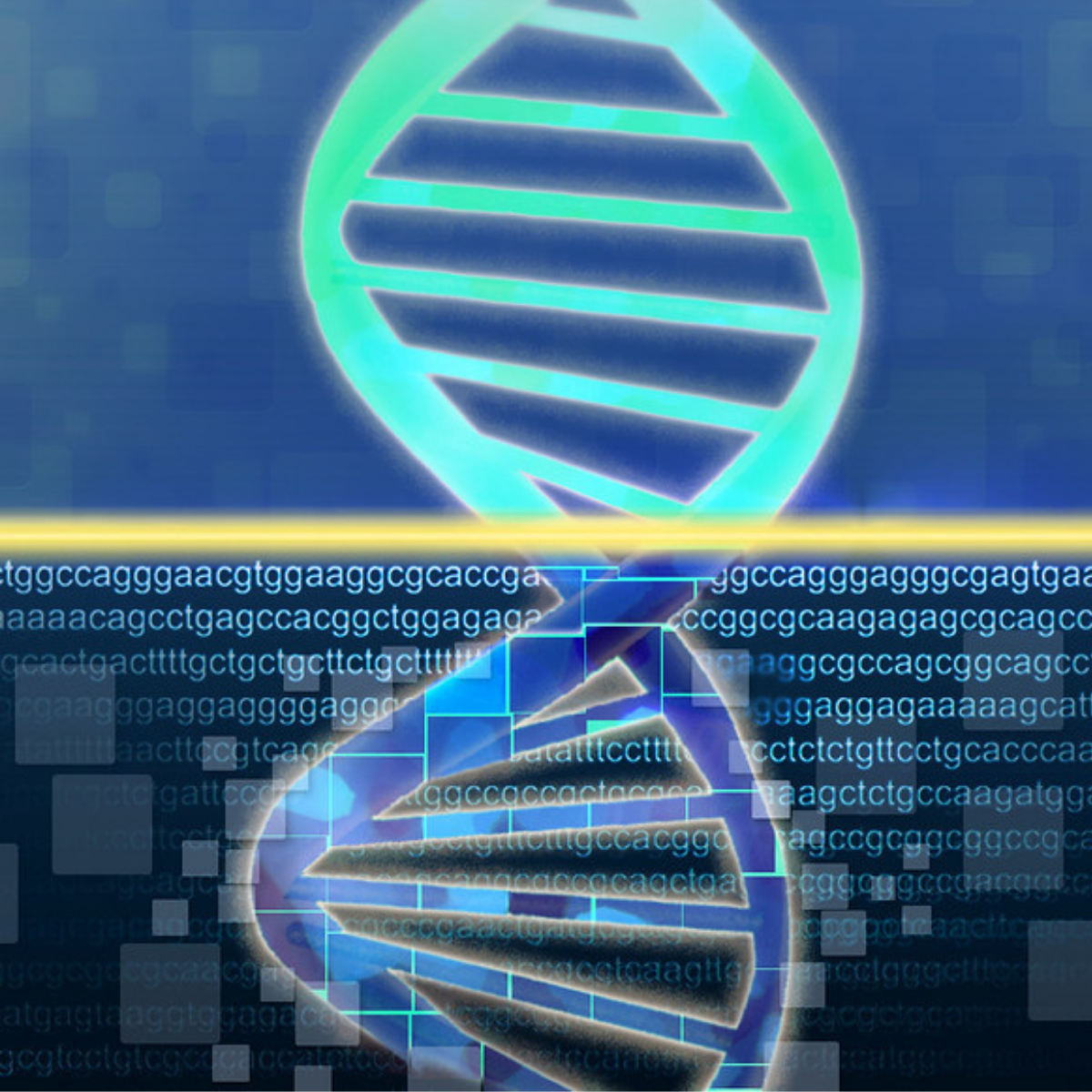
The future of DNA sequencing

Click here to listen to the full podcast episode
Despite the enormous advantages and potential of next-generation sequencing, science, as always, marches onwards. We are still creating new ways to read the book of life and push forward the frontiers of genetics.
One of the most recent advances is nanopore sequencing, which involves threading DNA through a tiny hole in a membrane called a nanopore. As each base passes through the membrane, a tiny electrical signal is generated. The signal is characteristic to each base, so detecting these minute electronic pulses means we can work out the sequence of the DNA passing through the membrane.
The technology reached the commercial market in 2014 when Oxford Nanopore released its MinION sequencer. For more on that story, listen to our previous episode, ‘Building an Army of MinIONs.’
Unlike next-generation sequencing technologies like Illumina’s, nanopore sequencing can analyse long sections of DNA, with no need to fragment the DNA before sequencing, and almost no sample preparation is required. Because no DNA is synthesised in the sequencing process, there’s no need to add nucleotides, enzymes or other chemical reagents. As a result, nanopore sequencing dramatically reduces the cost of sequencing and removes the need for a highly equipped laboratory.
Oxford Nanopore is now selling sequencers for less than $1000 that fit in the palm of your hand and plug into your computer, so you can analyse DNA quickly and cheaply, anywhere in the world. They’ve been a boon for researchers working out in the field, such as those tracking outbreaks of infectious diseases, including the current COVID-19 pandemic.
Other long read technologies are also coming through, such as PacBio’s SMRT sequencing technology, the brainchild of Jonas Korlach. Fascinated by the form and function of molecular ‘machines’ inside cells, Korlach started studying one of the most impressive of them all: DNA polymerase.
Aware of the DNA sequencing techniques at the time, which relied on DNA polymerase adding fluorescent nucleotides to large numbers of short fragments of DNA, he started to wonder if he could build a microscope that would enable him to spy directly on DNA polymerase as it added each coloured base to a single strand of DNA, creating an entirely new sequencing technique..
This wasn’t exactly a practical idea: the best microscopes at the time could visualise nothing smaller than a blob of 500 polymerase molecules - certainly not the single enzyme and DNA strand that Korlach wanted.
Luckily for him he was working in the lab of Watt Webb, who was an expert in high resolution imaging techniques. Together with Webb, and nanoengineers Steve Turner and Harold Craighead, Korlach eventually succeeded and by 2009 they had published the first iteration of their method.
At the heart of SMRT sequencing is a chip covered in millions of tiny holes. And by tiny I mean tiny - they’re so small that a single molecule of DNA and polymerase can be trapped in there, and they’re also so small that light can barely get through.
Fluorescently labelled bases are added to the chip - each of the four letters labelled with a different colour. As DNA polymerase gets to works copying the DNA strand and a new letter binds, it gives off a flash of coloured light, according to whichever base has just been added.
Like with Illumina sequencing, watching the pattern of flashes reveals the underlying sequence of the DNA strand in that spot. But unlike Illumina’s technology, SMRT sequencing can read much longer single strands of DNA, up to many thousands of letters at a time, scaled up millions of times on each chip. Again, there’s a video of how it works on the page for this podcast at Geneticsunzipped.com
Finally, the latest innovation in DNA sequencing takes us another leap forward.This time, instead of improving how we read DNA, scientists are changing where we do it.
In December 2020, researchers from MIT reported a method called ‘In-situ DNA sequencing’, which integrates sequencing with microscopy to show scientists not only the sequence of DNA but where it sits within the 3D structure of the cell nucleus.
The process involves fixing cells to a glass surface and then amplifying thousands of short segments of DNA at their original location using fluorescently labelled bases, each a different colour. The reaction is observed using fluorescence microscopy, giving the scientists a map of the sequences within the cell as they are amplified, pinpointing where they sit within chromosome structures.
Although it’s a brand new technique, advances that allow us to sequence DNA within cells could give us new opportunities to investigate how the 3D organisation of DNA affects its function and observe structural changes that are associated with ageing and diseases, including cancer and brain disorders.
As Fred Sanger said when he was awarded the Nobel Prize in 1980, “... [A] knowledge of sequences could contribute much to our understanding of living matter.”
The progress of DNA sequencing over the past few decades has been tremendous. But there is undoubtedly much more to come as we continue to learn how to read the book of life. In the words of Sanger’s co-Laureate Walter Gilbert, “I think the future of science will continue to astound us.”
References

